Computational Transport Oncophysics
This is an IAS project run in collaboration with IAS Hans Fischer Senior Fellow Prof. Bernhard Schrefler from the department of Civil, Environmental and Architectural Engineering of the University of Padua.
In the field of transport oncophysics cancer is regarded as a disease of multiscale mass transport deregulation involving biological barriers. Cancer physics is dominated by cell, particle and molecule transport through biological tissues. Furthermore, drug delivery involves transport through heterogeneous tumor and healthy tissue. In order to reduce the highly complex behaviour of tumors, physical models are necessary. One of the most advanced cancer models is the continuum model for tumor growth including angiogenesis developed by Prof. Schrefler and co-workers which is based on multiphase flow through a deformable porous medium. It consists of three fluid phases, i.e. tumor cells (TCs), divided into living and necrotic cells, healthy cells (HCs) and interstitial fluid (IF) and one solid phase, the extracellular matrix (ECM). In the current project, we aim to enhance this model with a patient-specific vasculature model by means of one-dimensional blood vessel models. Then, blood flow and transport of therapeutic agents in the multiphase tumor growth model and the vasculature model will be coupled consistently to make patient-specific predictions of the efficacy of treatment possible.
Components of the multiphase tumor growth model
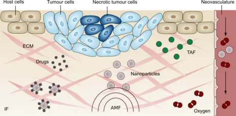
The model comprises a solid phase, the extracellular matrix (ECM), three fluid phases host cells, tumor cells and the interstitial fluid (IF), as well as the neovasculature which is modelled as an independent porous network. In addition, the phases transport species, namely necrotic tumor cells, oxygen, TAF and nanoparticles. The nanoparticles can either transport drugs to the tumor or be used to generate heat with an alternating magnetic field (AMF).
Growth of a Three-dimensional vascular tumor over 15 days
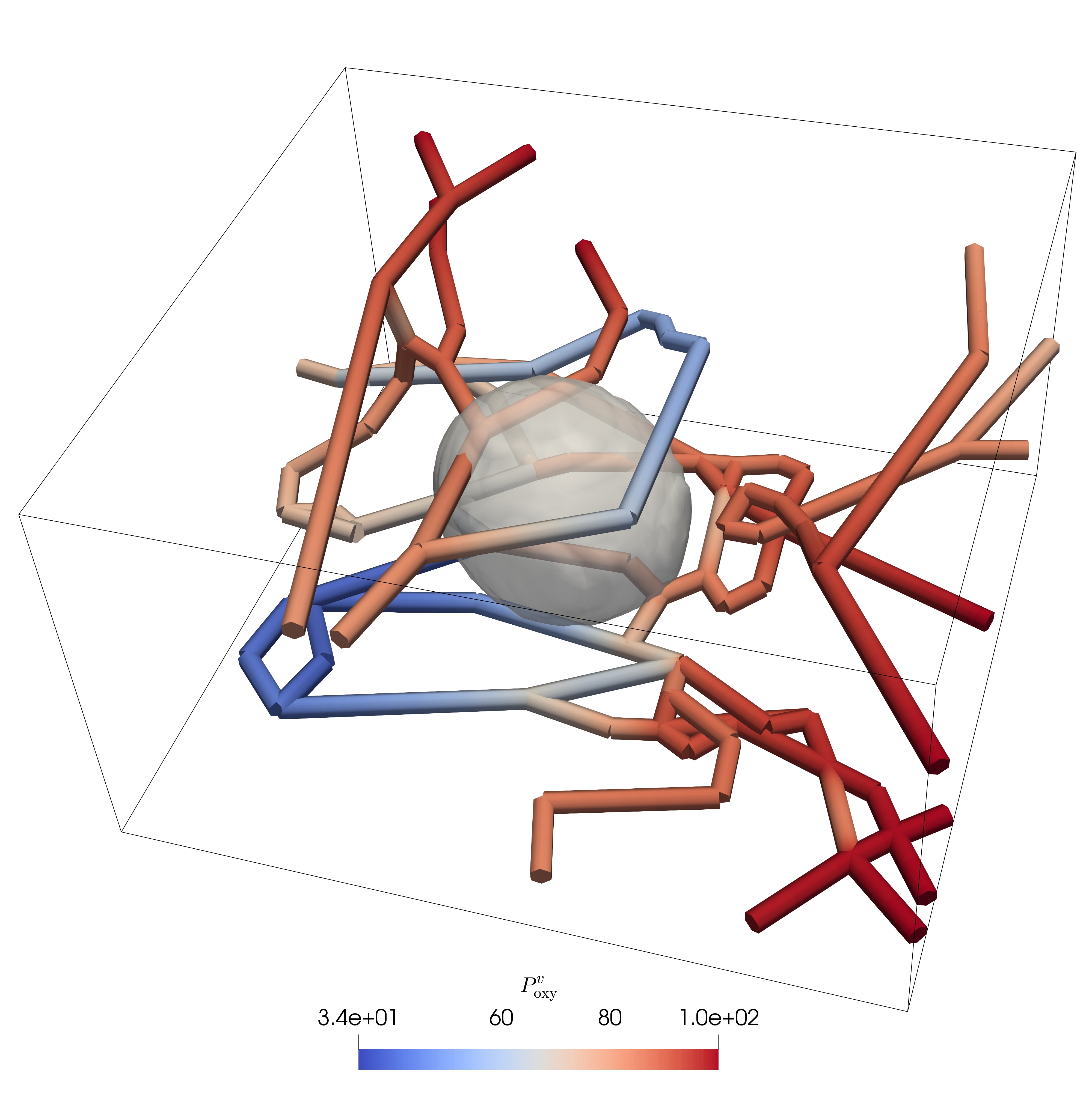
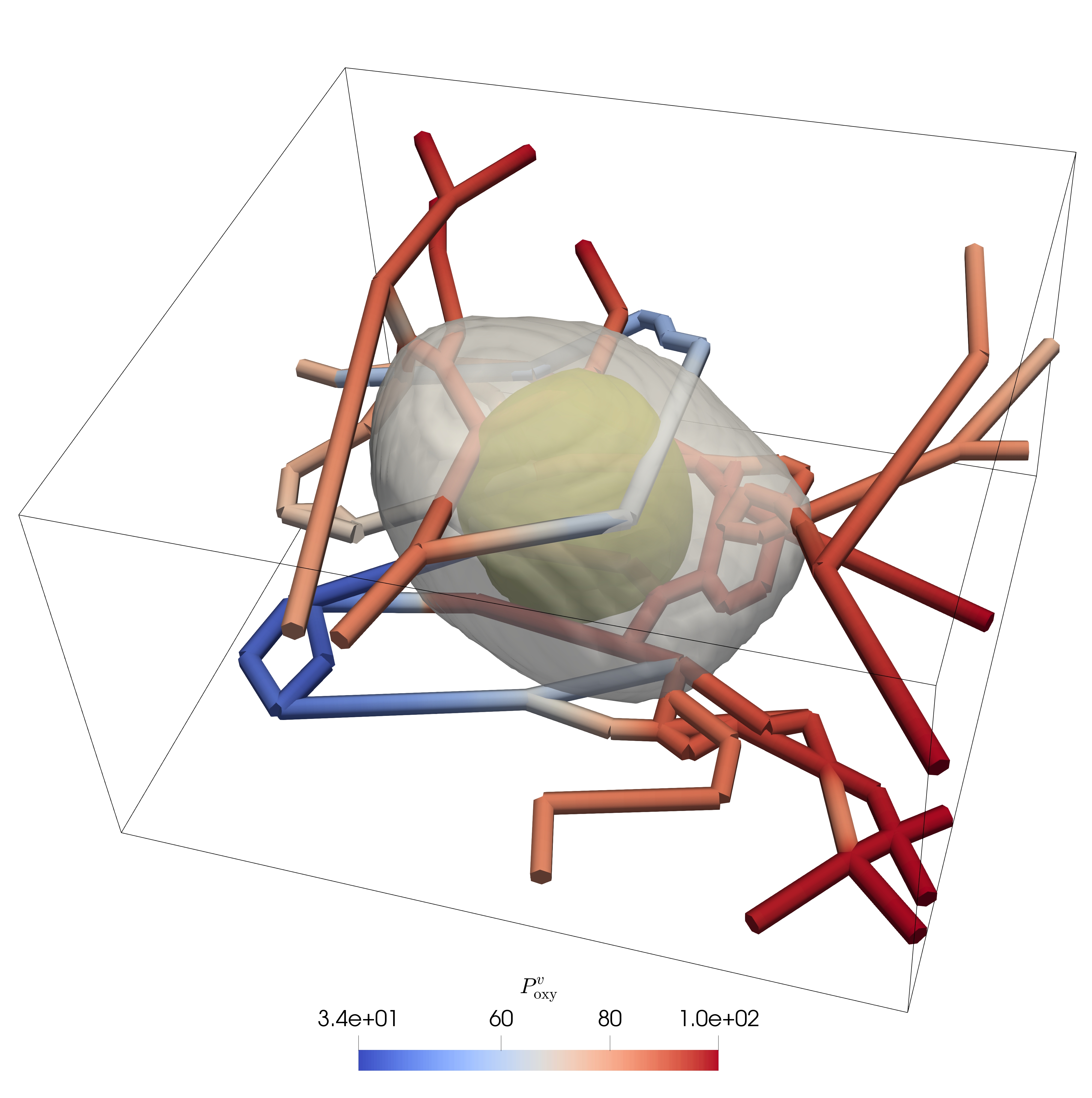
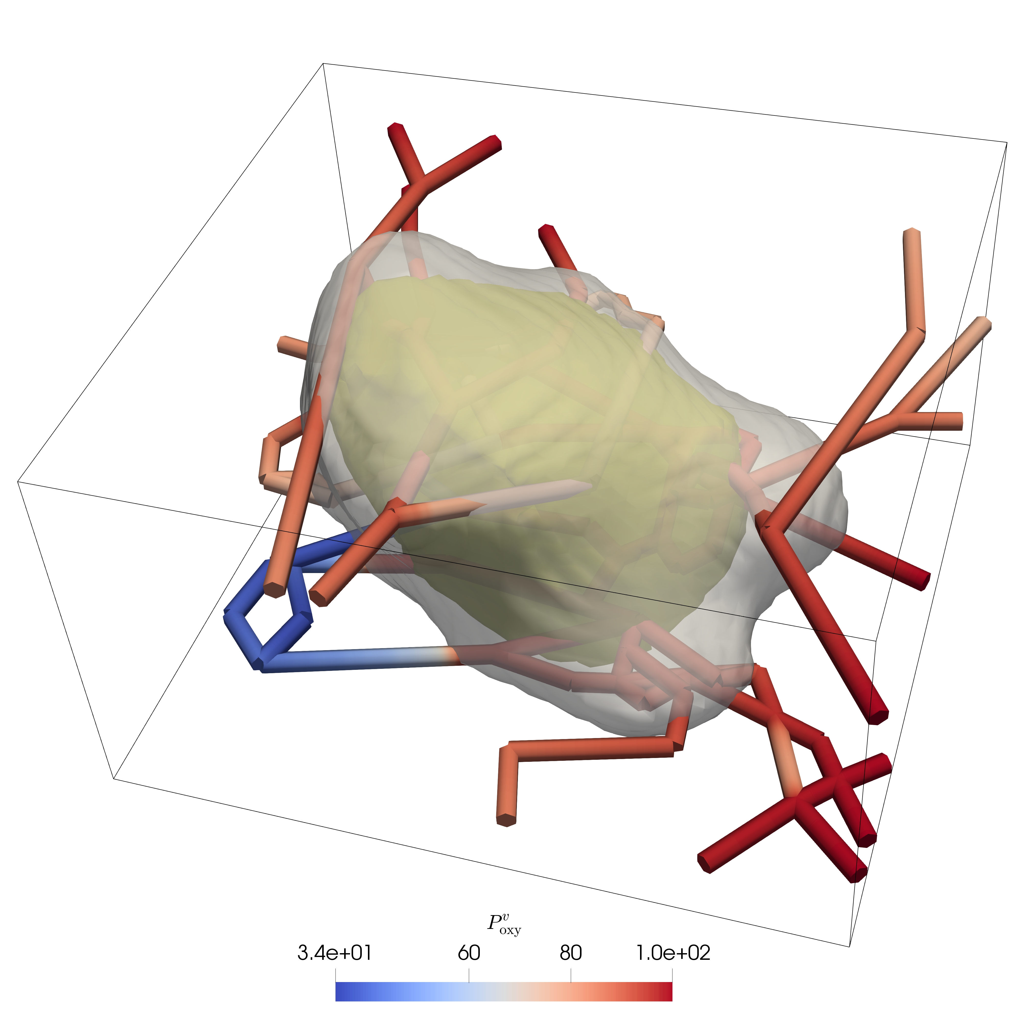
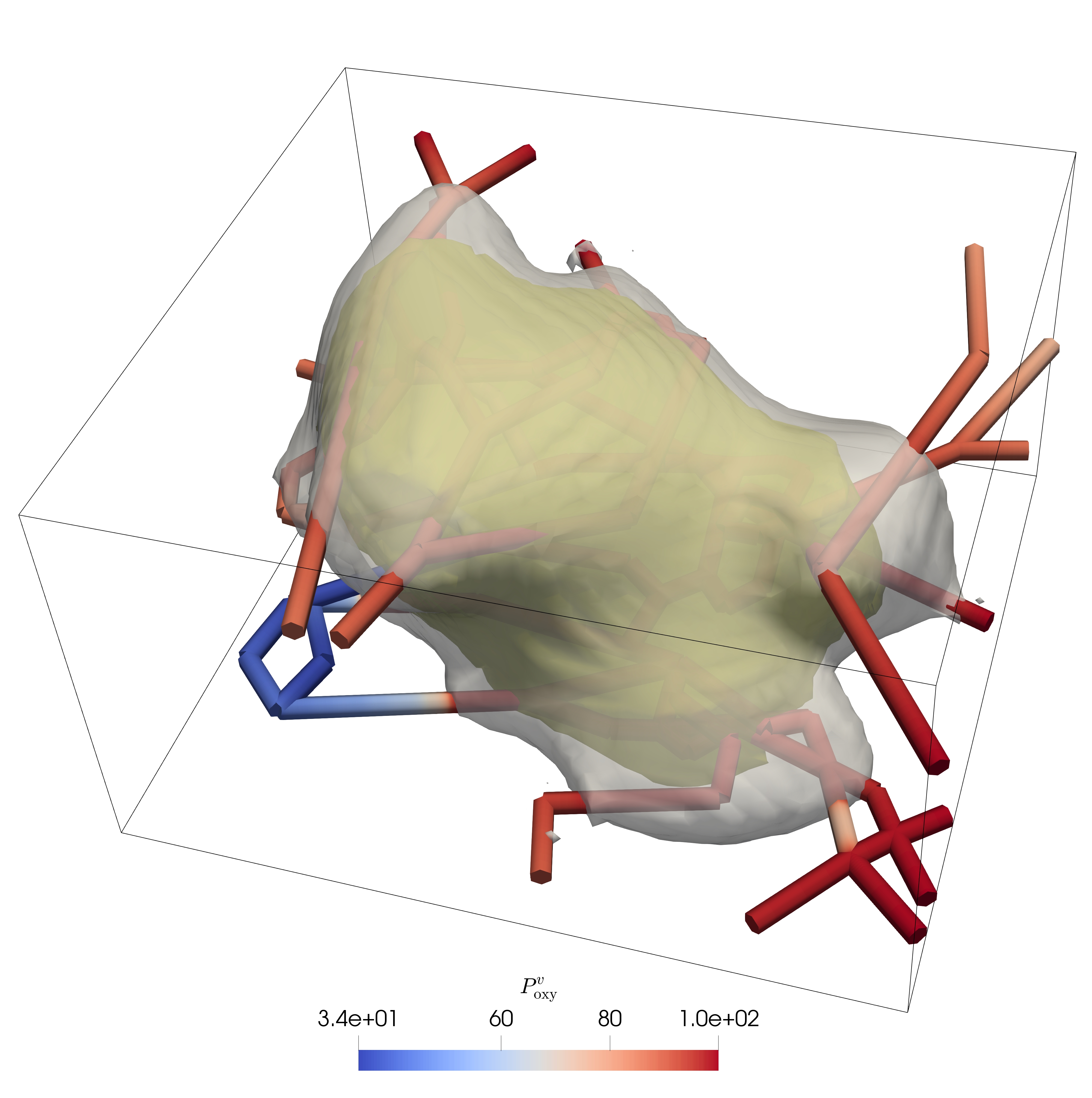
Three-dimensional growth of a tumor along a preexisting blood vessel network (outline of tumor visualized in grey, outline of necrotic core visualized in olive green)
Tumor follows an initial radial growth pattern. After several days, a necrotic core starts to appear in its interior since nutrient diffusion from the outside to the inside of the tumor is a limiting factor. Nevertheless, tumor growth can continue along the vasculature by co-option of the blood vessels because in this region oxygen concentration is highest.
Growth of a two-dimensional vascular tumor over 24 days
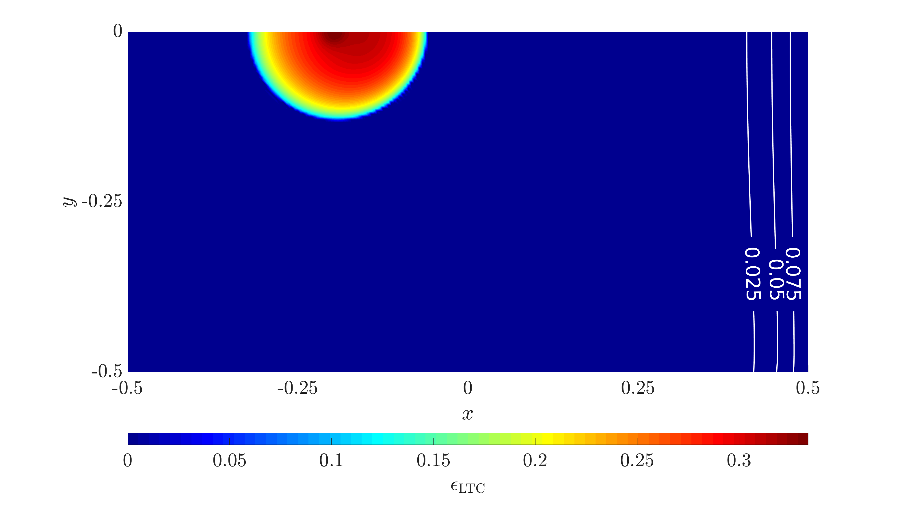
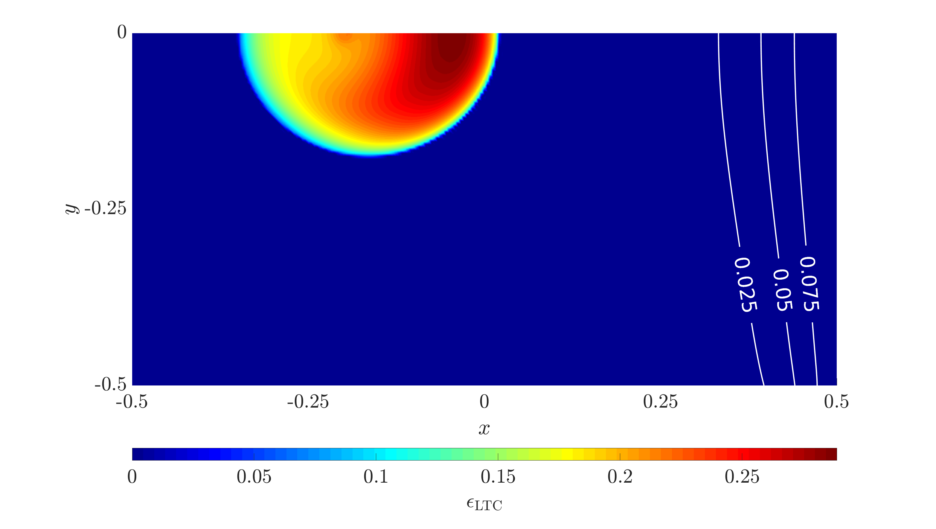
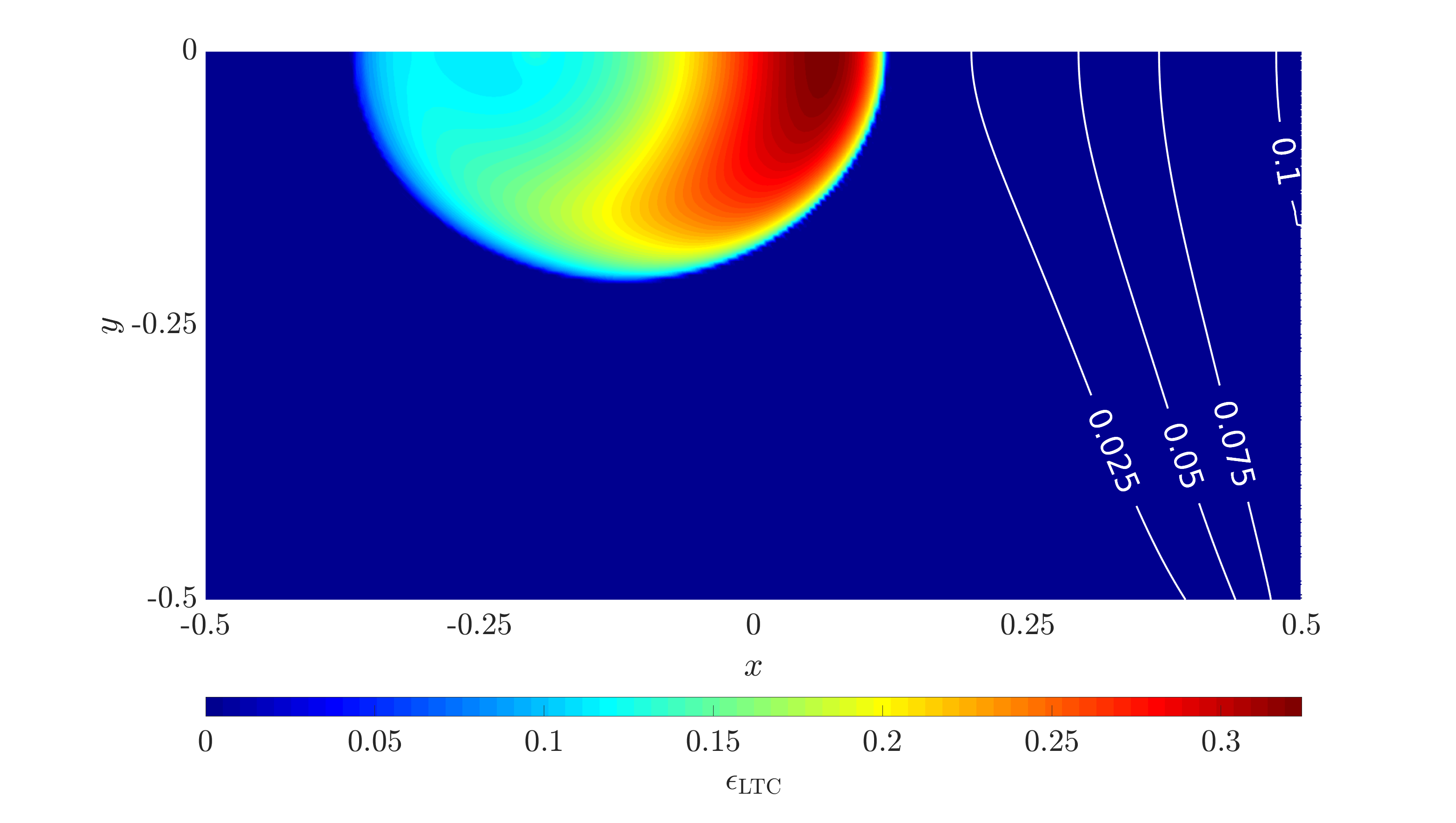
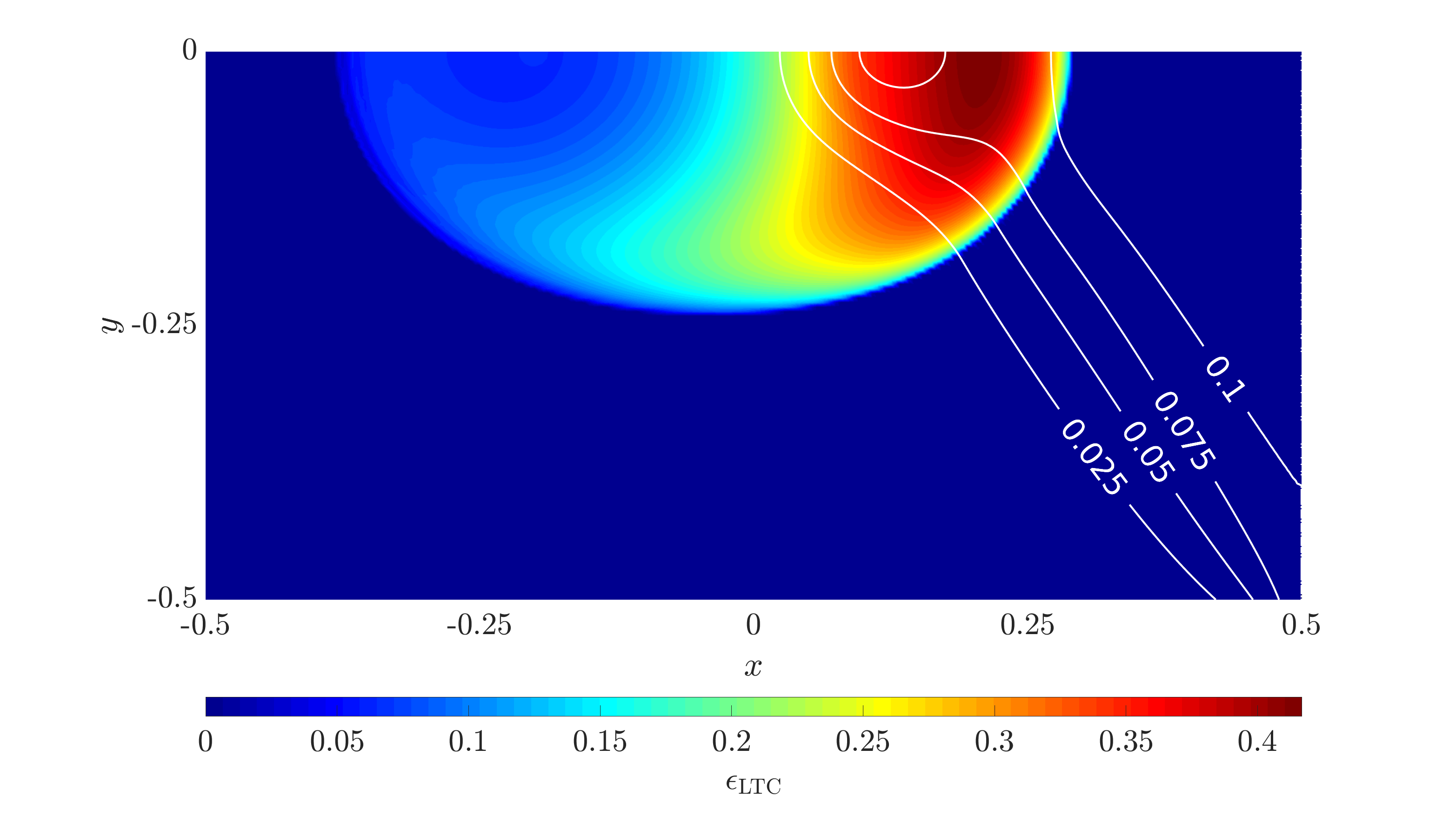
Evolution of living tumor cells and neovasculature (color scale denotes volume fraction of living tumor cells and contour lines neovasculature)
Tumor grows in half-moon shape towards the source of oxygen on the right. Tumor angiogenic factors are constantly produced by hypoxic tumor cells which triggers angiogenesis from the right.