Biomedical: Multiphysical & Multiscale Modeling of the Human Heart
Ph.D. students: Marc Hirschvogel, Julia Hörmann, Lasse Jagschies, Andreas Nagler, Martin Pfaller, Amadeus Gebauer
Postdoc: Stefanie Heyden
Faculty: Cristóbal Bertoglio, Michael Ortiz, Wolfgang Wall
We are currently developing a detailed, multiphysical computational model of the complete human heart (atrias and ventricles). We are working on the main aspects of the cardiac physics like electro-physiology, electromechanics, detailed fibre architecture, blood flow, valves and peripheral tissue. The project involves the collaboration with international research groups, the local industry and clinicians, and has been partially funded by the Institute for Advanced Study at TUM.
Geometry
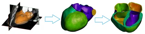
Medical image data is acquired either from Computed Tomography (CT) or Magnet Resonance Imaging (MRI). These raw images are transformed to the same coordinate system via image registration. Depending on the type (e.g. CT, cine MRI, morphological MRI, diffusion tensor MRI), the images are either segmented manually or semi-automatically. The resulting geometry is then exported as a surface mesh. Finally, a 3D volumetric finite element (FE) mesh is generated. In order to apply boundary conditions and construct the fiber organization, the mesh is further divided into different surfaces and volumes.
The following picture shows the geometry construction process.
Collaborators
Center for Mathematical Modeling, Universidad de Chile
Institute for Biomedical Engineering, ETH Zürich
German Heart Centre Munich, TU München
Department of Nuclear Medicine, TU München